
Pathophysiology of Dystonia
Dystonia is a movement disorder of yet unknown causes and pathophysiology. It is characterized by sustained or intermittent muscle contractions causing abnormal, often repetitive, movements, postures, or both.
Focal dystonias are the most common forms of dystonia and can affect different muscle groups (e.g., cervical dystonia, focal hand dystonia, laryngeal dystonia, blepharospasm). The treatment of this disorder is currently limited to symptom management, typically, with botulinum toxin injections into the affected muscles.
Our long-term goal is to identify the neural mechanisms underlying the pathophysiology of focal dystonia and to develop new strategies for enhanced clinical management of this disorder, including its accurate diagnosis, prediction in persons at-risk, and novel therapeutic strategies.
We use a variety of neuroimaging methodologies, including functional MRI (fMRI) and electroencephalography (EEG) for mapping of brain functional activity and networks, pharmacological fMRI (phfMRI) for the assessment of drug effects on brain function, high-resolution structural MRI and diffusion-weighted imaging (DWI) with tractography for evaluation of brain structural organization, and positron emission tomography (PET) with radiolabeled ligands for neuroreceptor mapping.
Imaging Genetics of Laryngeal Dystonia
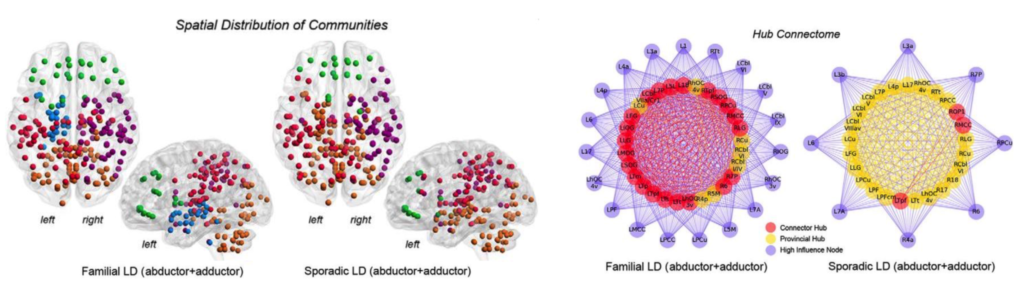
Laryngeal dystonia is isolated focal dystonia characterized by selective impairment of voluntary voice control predominantly during speech production. Despite well-characterized clinical features of LD, its causes and pathophysiology remain unclear. Consequently, the absence of objective biomarkers of LD leads to diagnostic inaccuracies, while the lack of understanding of its neural and molecular targets hinders the development of novel therapeutic opportunities for these patients. Funded by the National Institute on Deafness and other Communication Disorders, National Institutes of Health (NIDCD/NIH R01DC011805), our research program is set to identify imaging and genetic biomarkers of LD development and manifestation. We use a comprehensive approach of multi-modal neuroimaging, machine learning, and next-generation DNA sequencing as tools for the discovery of the mediating neural mechanisms that bridge the gap between the DNA sequence and LD pathophysiology. Knowledge obtained from these studies is expected to have a direct clinical impact by establishing enhanced criteria for accurate and objective diagnosis, screening of persons at-risk, and evaluation of mechanism-based novel pharmacological and/or surgical therapies for these patients.
Brain Networks in Dystonia
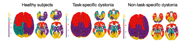
Task-specific focal dystonias are characterized by selective activation of dystonic movements during the performance of highly learned motor tasks, such as writing or speaking. To date, we have only limited knowledge about the distinct neural abnormalities that lead to the development of task-specificity in focal dystonias, which affect similar muscle groups but result in different clinical manifestations, such as writer’s cramp vs. pianist’s dystonia or spasmodic dysphonia vs. singer’s dystonia. Funded by the National Institute of Neurological Disorders and Stroke, National Institutes of Health (NINDS/NIH R01NS088160), our goal is to dissect the pathophysiological mechanisms underlying the phenomenon of task specificity in isolated focal dystonias using multi-level brain network analysis in conjunction with neuropathological examination of postmortem brain tissue from patients with dystonia. Rather than viewing these disorders as interesting curiosities, understanding the biology of task-specific activation of motor programs is central to understanding dystonia.
Neurotransmission in Dystonia
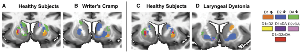
Despite the recent progress in elucidating functional brain abnormalities within the basal ganglia-thalamo-cortical circuitry in focal dystonias, there is a fundamental gap in understanding the neurochemical correlates underpinning the functional alterations in these disorders. Our goal is to provide detailed knowledge about the neurotransmission via GABAA, D1– and D2-family receptors in patients with different forms of focal dystonia. This information will help determine the contribution of GABAergic and dopaminergic neurotransmission to the pathophysiology of dystonia, as well as identify potential new pharmacological targets for novel treatment options.